Articles

Young Adults Male Infertility Causes & Treatment

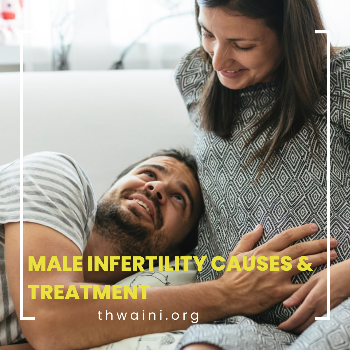
Infertility is a common problem affecting young adults. At least one and six couples would be suffering from this condition. It is the inability to conceive after one year of unprotected intercourse. While traditionally fingers are always pointing towards the female partner, men are equally responsible for inFertility with 50% being caused by the male factor.

Of course, there are other factors causing infertility despite Both partners having normal parameters. When it comes to male factor infertility, there are several causes such as infections, trauma exposure to toxins, or congenital problems. Many of these factors are easily treatable, however, some might require an intervention to facilitate conception (Assisted fertilization). While this started fairly recently in the 80s with limited success, With the advancement of technology and medical sciences, success rates of dramatically increased.

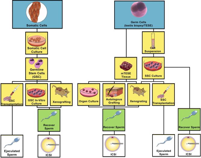
Sperm harvest, though challenging, is becoming more feasible with the improvement in technology. This includes Microscopic testicular sperm extraction, Epididymal sperm aspiration, etc. The application of regenerative medicine in many aspects of healthcare is becoming more popular, however, when treating infertility, this remains to be experimental. Watch the space!

Dr. Ali Thwaini is one of the urologists and provides male infertility treatment in Dubai

Blood in the urine Have you ever noticed that the color of your urine is darker than normal?

Blood in the urine (hematuria) could manifest itself from a variety of conditions. This could be quite alarming finding when it happens. People would normally Google their symptoms and Dr. Google gives them the scare of their lives when going through the list of causes! First, red-colored urine doesn’t automatically mean that there’s blood in it.

There are various conditions that might lead to darker color urine, sometimes red, but aren’t associated with bleeding; these could be simply dehydration, excessive beetroot intake, some medicine ( e.g. Rifampicin), or excess intake of certain vitamins. When you have altered color urine, the best thing is to get it checked with the laboratory.

Urine is normally tested using a special dipstick test that contains chemicals able to detect blood constituents. This is a very fast test, cheap, and fairly reliable. Urine microscopy is another alternative. Both are non invasive and easily performed. If these tests confirm the present of blood, further assessments are required. These are in the form of radiological imaging ( ultrasound scan, or sometimes CT scan).

The other test is a direct visual inspection of the urethra and urinary bladder. This is carried out in the clinic using a flexible cystoscope under local anesthesia. It’s a very common, easy, and efficient procedure to confirm, or better, rule out urinary bladder pathology. Whilst hematuria is an alarming finding, mostly it’s due to non-sinister causes; these could range from urinary tract stones, infections, benign enlargement of the prostate. However, occasionally, there could be a significant underlying pathology, such as urinary tract cancer. There are some predisposing factors though; with the main being smoking. And yes, Shisha smoking, though the thought of as a benign social hobby, is by no means less harmful than cigarette smoking (one shisha smoking equals 70 cigarettes!).

Therefore, if you develop such a symptom, it is always better to visit your urologist to have the necessary tests carried out. All these tests are available at the Mediclinic City Hospital.

Renal cancer The silent killer

Renal Cancer is one of the come in urological counters affecting people are different stages of their lives. There are about 75,000 renal cancer new cases diagnose every year the United States Constitution saying about 5% of all other cancers and women are equally affected. While there are no definite predisposing factors to the development of renal cancers, There is a weak link between obesity and hypertension and, of course, smoking, to the development of real Cancers.

Most are diagnosed Anecdotally, however, some Are hereditary. Such as VHL disease. Before the advancements in medicine and surgery, kidney cancers were diagnosed rather late and patients were beyond the stage of cure. Due to the rightfully low thresholds and imaging, most of these cancers tend to be diagnosed incidentally and in early stages, Hence the name “incidentalomas”.

The benefits of early diagnosis of kidney cancer Are multifactorial; With early diagnosis, Tumors tend to be at a very early stage And patients are being offered kidney Sparing operations. This type of treatment has the potential of preventing The development of chronic kidney disease. Also, when the tumors are diagnosed at an early stage, especially in people who are high-risk surgical candidates, there is the option of minimally invasive treatment, in the form of the use of high or low temperatures to remove the cancer cells; this is in the form of radiofrequency ablation, Microwave ablation, And cryoablation. These methods have safe, proving their efficacy and safety.

The third benefit of diagnosing early kidney cancer is having a chance to follow them up closely In order to understand the biology of the disease. Several studies have shown that small kidney cancers tend to grow at a very slow pace the elderly and there is an option of active surveillance in this particular cohort of patients. Having said all the above, nephron-sparing surgery remains the gold standard in early localized kidney cancers, especially when technically feasible and safe. Surgery, on par with Medicine, has significantly progressed, and we have moved from open surgery to laparoscopic and then robotic surgery. While all options have proven their Efficacy in curing kidney cancer, the latter two surgical options have proven their safety and efficacy in providing an early return to normal function with less perioperative complications.

At Mediclinic and City Hospital, all the above modalities are available in treating kidney cancers and Patient’s are well managed by the multidisciplinary team to make their journey through illness and recovery as short and smooth as possible.

Festive Seasons and their Effects on your Urinary System

Christmas has just passed and we are still in the partying mode, preparing for the new year. It’s been tough couple of years with yet another wave, albeit less virulent, of COVID’s new disguise; Omicron.

We are on the verge of winning our war against it. It’s indeed worth the celebration. One has to be careful though with the effects of alcohol on our urinary system. The Celebration should not be at the expense of our health and the safety of people around us. Here are some facts about alcohol and its effects on our urinary system.

The effect of Alcohol on the kidneys:

Alcohol alters the filtration function of the kidneys adversely resulting in the reduction of its efficacy, thus rendering them less able to filter the blood.

Alcohol also affects the ability to regulate fluid and electrolytes in the body. It has a natural diuretic effect leading to frequent visits to the toilet after many drinks. This, however, leads to intracellular dehydration, resulting in loss of excess fluid and an Increase in electrolyte concentration in the body.

Regular heavy drinking has been found to double the risk of chronic kidney disease, which does not go away over time. Even a higher risk of kidney problems has been found for heavy drinkers who also smoke.

The Centers for Disease Control estimates that most American adults (two out of three) drink alcohol. Too often, some of these regular drinkers have more than five drinks at one time. In fact, about a quarter of drinkers reported they had done this on at least one day in the past year. “Binge” drinking is even worse. It has harmful effects on the kidney that can even lead to acute kidney failure. A sudden drop in kidney function is called acute kidney failure. This often goes away after a time, but it can occasionally lead to lasting kidney damage.

Added to that is the effect of chronic drinking on the increase in blood pressure resulting in different levels of kidney damage with protein loss in the urine.

Studies have demonstrated the effects of chronic alcohol intake on the cellular structure of the building brick of the kidney; The nephron. It’s been shown that the basement membrane of various aspects of the nephron develops an increase in its thickness, therefore hindering its ability to perform its filtration and concentration functions properly.

The effects of alcohol on the urinary bladder:

Alcohol has a double adverse effect on the urinary bladder: It has a strong diuretic affect leading to excessive frequency and urgency.

It will also lead to dehydration resulting in more concentrated urine, which causes burning of micturition by the concentrated urine.

It has been shown previously by various experiments when the bladder gets distended, the intravesical pressure increases to a peek after which (especially in situations not permitting paying visits to the toilet) the pressure drops down simply due to the thinning of the bladder muscle. This results and the reduction and pressure necessary to generate in order to empty the urinary bladder (law of Laplas).

Also, chronic alcohol intake leads to nerve damage resulting in “alcoholic cystopathy”. This would potentially lead to the current urinary retention.

In summary:

Drinking can be fun in certain cultures, let’s be honest. Excessive alcohol intake can lead to injury, accidents, serious embarrassment and long-term health problems. Even drinking small amounts of alcohol increases your cancer risk.

There are a few tips for those who would enjoy having a drink with their family and friends.

  • Eat before drinking to minimize the direct effect of alcohol on your body.
  • Drink plenty of water.
  • Don’t mix alcohol with sugary or energy drinks.
  • Avoid salty snacks – they will make you thirsty and likely to drink more.
  • Be in control of the number of drinks you take: Set yourself a drinks limit and stick to it. Avoid drinking in rounds (especially with friends who drink too much). Try to finish your drink before you start another, rather than topping up your glass.
  • Slow down when you drink: To keep safe, slow down your drinking to 1 drink per hour. You can do this by:
  • drinking non-alcoholic drinks as well as alcoholic drinks
  • drinking water to quench your thirst before you start drinking alcohol
  • opting for low-alcohol drinks. sipping rather than gulping

Wish you a Happy New Year

References:

Sexually Transmitted Diseases

Sexually transmitted diseases (STDs), or sexually transmitted infections (STIs), are contagious diseases that are passed between persons through direct sexual contact, through vaginal, oral, and anal sex. But sometimes they can spread through direct skin contact. This is because some STDs, like herpes and HPV, are spread by skin-to-skin contact.

There are several types of STDs that can be caused by bacteria, viruses, and parasites, including

  • Chlamydia
  • Genital herpes
  • Gonorrhea
  • HIV/AIDS
  • HPV
  • Pubic lice
  • Syphilis
  • Trichomoniasis

Who is affected by sexually transmitted diseases (STDs)?

Most STDs affect both sexes, but in many cases, the health problems they cause can be more severe for women. If a pregnant woman has an STD, it can cause serious health problems for the baby.

Symptoms of sexually transmitted diseases:

STDs might cause minimal or even no symptoms. So it is possible to have an infection and not know it. Yet they can still be passed on to others.

Symptoms include

  • Unusual discharge from the penis or vagina
  • Sores or warts on the genital area
  • Painful or frequent urination
  • Itching and redness in the genital area
  • Blisters or sores in or around the mouth
  • Abnormal vaginal odor
  • Anal itching, soreness, or bleeding
  • Abdominal pain
  • Fever

Diagnosis:

If you are sexually active, you should talk to your health care provider about your risk for STDs and whether you need to be tested. This is especially important since many STDs do not usually cause symptoms.

Some STDs may be diagnosed during a physical exam or through microscopic examination of a sore or fluid swabbed from the vagina, penis, or anus. Blood tests can diagnose other types of STDs.

Treatment for sexually transmitted diseases:

Antibiotics can treat STDs caused by bacteria or parasites. There is no cure for STDs caused by viruses, but medicines can often help with the symptoms and lower your risk of spreading the infection.

Prevention of sexually transmitted diseases:

Attempts to minimize direct genitalia and skin contact via the usage of latex condoms greatly reduce but do not completely eliminate the risk of catching or spreading STDs. The most reliable way to avoid infection is to not have anal, vaginal, or oral sex.

There are vaccines to prevent HPV and hepatitis B.

Is it safe for people with UTI to keep fasting during Ramadan?

The holy Ramadan season is upon us and we are devoted and hardcore dedicated for the long ritualistic fasting hours. To make this Ramadan season safe and healthy, urologist Dr. Ali Thwaini has a lot to share!

With long fasting hours, it is obvious that the water intake lowers to below the sufficiency levels and it is a major concern. Why?

With scorching sun, unbearable heat waves, and longer days, dehydration is a common issue accompanied by fatigue and weakness. This can ultimately lead to kidney stones and Urinary Tract Infections (UTI).

With dehydration clutching your body, dry skin and mouth, constipation, severe headache, thirst can be the resultant issues to resolve.

Kidney, Ureter or Bladder (KUB) stones and UTIs can arise in people who do not care to have at least 8 to 10 glasses of water a day. This is because, with no adequate intake of water, dilution of uric acid is not properly done and pH levels drastically reduce, leading to soaring acid levels and kidney stones start forming in your KUB.

UTIs tend to be a commonly sighted problem during fasting, especially in women.

How do you find you are afflicted with UTI?

The initial symptoms of UTIs

  • Having a burning sensation while urinating.
  • Severe pain in the lower abdomen.
  • Spotting blood in the urine.
  • Lower back pain.
  • Frequent urination accompanied by pain.

How UTIs affect your bladder can be put in simple words, it’s when there is an infestation of bacteria within the bladder. And this happens generally due to 2 main reasons:

  • Reduced intake of water, hence the bacteria are not flushed out of the bladder and hence start infesting.
  • Constipation can also result in the building up of bacteria which can resultantly affect one’s KUB.

It’s a major deduction that people with Diabetes are susceptible to bacterial, viral, and fungal diseases. Without proper medication, uncontrolled diabetes can result in chronic renal disease.

Quick remedies

  • Take special concerns to stay highly hydrated during the nonfasting hours.
  • Make sure to maintain good glycemic control with a proper diet plan and antidiabetic therapeutics.
  • Drink at least 3 liters of water a day and stick to a low carb, low protein, and less salted diet
  • Drink as much as fluids during Iftar and Suhoor in the form of fresh fruit juice to boost your immunity and nourish your body with a plethora of vitamins and minerals.
  • Skip too many cups of coffee, since it’s an infamous diuretic and can dehydrate your entire system in the blink of an eye.
  • Consume lime water with less added sugar, to boost the citrate levels of your body.
  • Intake cranberry juice is also highly recommended to keep UTIs at bay.

With a proper diet plan and a hydrated body system, the majority of UTIs can be avoided to an extent. However, it’s wise to consult a urologist, for people affected by kidney disease, kidney stones, or UTIs before fasting to keep things safe and healthy!

Undescended Testicles: Symptoms, Diagnosis & Treatment – Dr.Ali Thwaini Urologist Dubai

Undescended Testicles

This is a rather common condition and is even more common in premature babies. Around one in 20 male babies is born with an undescended testicle. In about one in 70 cases, the testicle remains undescended when the child’s testicles are not in their usual place in the scrotum. Generally, only one of the testicles is affected, but on rare occasions, both testicles fail to travel to the scrotum. 

Towards the end of pregnancy, the testicles travel through a passage into the scrotum. Both testicles should be in the scrotum by the time the child is one year old.

In some children, the testicles may be in the scrotum for much of the time, but cannot be felt there because they naturally rise back into the body through fear or cold temperatures. A parent can usually find this out by putting the child in a warm bath and checking whether they can feel both testicles. If this is the case, there is no cause for concern.

Symptoms of undescended Testicles

The condition is asymptomatic for the child but the affected side cannot be felt in the scrotum. The child will not be in pain, and the undescended testicles will not interfere with any bodily function.

However, if one of the testicles becomes twisted (testicular torsion), this will be painful, either in the groin area or the abdomen, depending on the location of the testicle at the time.

Diagnosis of undescended Testicles

The mainstay of the diagnosis is by clinical examination, preferably in a warm environment in order to relax the scrofulous and allow maximum change to have a proper clinical assessment.

Aetiology

On rare occasions, the testicle does not descend due to other problems with the testicles themselves or with the male hormones. We do not know exactly why this happens, but it is not due to anything that happened.

Undescended Testicles Treatment

The method of treatment depends on the suspected cause. If the doctors suspect the testicles have not descended due to a hormone problem, they may suggest a short course of a hormone called human chorionic gonadotrophin (hCG). This is more likely to be suspected if neither testicle has descended.

If the doctor does not suspect a hormone problem, or if the testicles remain in the abdomen after the hormone treatment, the child will need a short operation under a general anesthetic called an orchidopexy.

Undescended testicles are best treated in early childhood, usually just before or around one year of age. The child’s testicles will need treatment as they do not seem to mature properly if left in the abdomen.

The amount of sperm and fertility levels seem lower in men who have had undescended testicles, and even lower if they were not treated early in childhood. This is because the testicles need to be a few degrees cooler than the rest of the body to produce sperm.

Children with undescended testicles have a higher risk of testicular cancer in the future. It is easier to check the testicles if they are in the scrotum. If the testicles remain in the abdomen or high up in the groin, this also increases the risk of testicular torsion.

What is an orchidopexy?

This is an operation to bring the testicles down from the abdomen to their usual place in the scrotum. This is a short operation under general anaesthetic, lasting about 45 minutes. Sometimes the operation needs to be done in two stages about six months apart.

In many cases, this can be as day surgery – the child will arrive at the hospital, have the operation and be able to go on the same day. Occasionally, a child will need to stay in hospital overnight.

Penoplasty (male enhancement)


The penile enlargement procedure is seldom discussed. Men in general are private about their privates at the best of times, and when it comes to such an intimate matter, they are even more introvert.


UTI in Pregnancy Dr.Ali Thwaini Urologist Dubai

UTIs during pregnancy are not uncommon and increase the risk of developing pyelonephritis, which is associated with an increased risk of fetal loss, premature delivery, and low birth weight babies. Screening can reduce the risk of this.

All women should be screened for asymptomatic bacteriuria at the 1st antenatal appointment

Symptomatic bacteriuria occurs in 17-20% of pregnancies. There are pathophysiological grounds to support a link to pre-labour,

premature rupture of membranes (PROM) and pre-term labour. Untreated upper UTI in pregnancy also carries risks of morbidity and rarely mortality to the pregnant women 

Physiological changes in the pregnant woman make her more likely to suffer both asymptomatic bacteriuria (AB), and urinary infection (cystitis, pyelonephritis).

2-9% of women are bacteriuric in the first trimester. 10-30% of women with bacteriuria in the first trimester develop upper urinary tract infection in the second or third trimester

High fever, whether caused by UTI or other infection, is associated with foetal loss, at any stage in pregnancy.

Benefits of screening for asymptomatic bacteriuria:

Early screening for and treatment of asymptomatic bacteriuria in pregnancy has maternal and foetal benefits.

A Cochrane review of 14 randomized trials of asymptomatic bacteriuria in pregnant women compared the antibacterial therapy to that with placebo or no treatment. The Cochrane review showed that antibacterial therapy was significantly more likely to clear asymptomatic bacteriuria, to lower the incidence of pyelonephritis, and to reduce the rate of preterm delivery or low birth weight babies.

Screening for asymptomatic bacteriuria in pregnancy

All women should be screened once for asymptomatic bacteriuria at the

1st antenatal (booking) appointment (NICE recommendation).

Do this by sending an MSU. DO NOT USE DIPSTICKS: they are not sufficiently sensitive.

If positive result, repeat as indicated in the flow chart over the page to ensure first test is reliable , as contamination can occur.

Managing symptomatic bacteriuria

Symptomatic bacteriuria in pregnancy should be treated (see over page for guidance on antimicrobials).

Use near-patient testing with dipsticks to assess the likelihood of UTI. Send urine for culture before starting empirical therapy. Send a repeat sample 7 days after completing treatment as a test of cure.

Antimicrobials for bacteriuria in pregnancy

The choice of antibacterial and the duration of therapy depend on a number of considerations:

  1. The relative contraindications to some antimicrobials in pregnant women (always refer to the BNF)
  2. Resistance of the organisms;
  3. Adverse effect profiles (including propensity to cause C. difficile-
  4. infection).
  5. Use MSU results, when available, to guide therapy even if this entails a change of empirical therapy.
  6. Since most antimicrobials are concentrated in urine, oral therapy is sufficient in most patients

Managing incidentally-found group B streptococcus infection in urine

The antenatal care service should be informed when a group B streptococcus (GBS), Streptococcus agalactiae, is isolated in urine. Women with GBS bacteriuria identified during the current pregnancy should be offered IV antimicrobial prophylaxis during delivery .

GBS bacteriuria, is associated with a higher risk of choriamnitis and neonatal disease. However, it is currently not possible to accurately quantify these increased risks.

Women with GBS urinary tract infection during pregnancy should also receive appropriate treatment at the time of diagnosis as well as IV prophylactic antimicrobials as the time of delivery. Treatment of GBS UTI during pregnancy should be treated as per culture sensitivities. Refer to BNF for further advice on appropriate antimicrobials during pregnancy.

Urine Sampling

The specimen should be mid-stream. Cleansing with water and holding the labia apart are not essential. Use of antiseptics for cleaning the perineum is not

recommended as this can cause false negative culture results. Refrigerate specimens to prevent bacterial overgrowth.

Interpreting a culture result:

The following usually indicates UTI in a patient with urinary symptoms. Higher counts have even higher positive predictive values:

  1. Single organisms ≥ 104 colony forming units (CFUs)/ mL
  2. Mixed growths’ indicates perineal contamination which reduces the significance of the culture. If a culture is still required, an MSU should be repeated with patient counselled on correct sampling technique
  3. Culture results should be interpreted in the light of near-patient dipstick testing.

Microscopy:

Microscopy is not available for the diagnosis of UTI except in children <3years to comply with NICE guidelines. Use near-patient testing with dipsticks to assess the likelihood of UTI, they are as sensitive and specific as microscopy for predicting the presence of infection. Urine microscopy is only performed for? glomerulonephritis, SLE, endocarditis, haematuria, casts, crystals, candiduria and Schistosomiasis and must be specifically requested with the relevant clinical details.

Treatment: please refer to the following chart:


Stem Cell Therapy in Male Factor Infertility: Research Proposal:

Background:

More than 10% of couples in the world experience fertility problems. Infertility, defined as failure to conceive a clinically detectable pregnancy after >12 months of unprotected intercourse, is a common condition, reported by 1 in 6 couples.

Stem cells exist as undifferentiated cells. They are present in the embryonic and adult stages of life and are considered as a source for differentiated cells that make up the building blocks of tissue and organs.

Due to their abundant source and high differentiation potential, stem cells are considered as potential new therapeutic agents for the treatment of infertility. Stem cells could be stimulated in vitro to multiply and then are utilized in vivo to “awaken’ the dormant spermatogonia” and are theoretically a potential source to develop various numbers of specialized cells including male and female gametes suggesting their potential use in reproductive medicine. During the past few years, considerable progress in the derivation of male germ cells from pluripotent stem cells has been made. In addition, stem cell-based strategies for ovarian regeneration and oocyte production have been proposed as future clinical therapies for treating infertility in women.

There are several sources for stem cells: Embryonic-derived stem cells (ESC), extra-embryonic derived SC, and mesenchymal derived SC. Each has their advantages and disadvantages. In this contest of male factor infertility, another source of stem cells could exit, albeit, a sparse source; this is derived from autologous Spermatogonial stem cells. Whilst harvesting them is a relatively simple procedure at an outpatient setting, and there are no ethical nor moral issues with harvesting them, they are relatively small numbers in the testis; hence, extracting them would be rather difficult and it is challenging to be maintained in culture. Moreover, there is a risk of immune rejection.

Challenges with stem cell therapy in male factor infertility:

Besides genetic factors, azoospermia also occurs due to injuries, exposure to toxicants, immune-suppressive and anticancer treatments. However, a large proportion of infertile males are diagnosed as idiopathic with unknown causes, reflecting poor understanding of the mechanisms regulating spermatogenesis and sperm function in humans.
While several sources exit to cultivate and improve stem cells for particular functions, generating pluripotent stem cells that have the potential to differentiate and undergo mitosis followed by meiosis into haploid cells remains challenging.
There have been few animal studies (particularly mice) where autologous mouse induced plutipotent cells have been produced (miPSC). Those actually were cultivated into mature spermatogonia and early spermatids. However, only a few studies made successful in producing spermatozoa in mice. This is yet to extrapolate and translate into human trials.

However, there are few human trials where a successful culture of hiPSC was achieved, resulting in spermatogonia and few spermatids.

Possible sources of stem cells:

These can be derived from either embryo of the placenta (the former might have ethical problems and both might be associated with future, though minor, risks of mutations). Other cells are derived from adult bone marrow and fat cells, which are rich in stem cells.
The function of stem cells in male factor infertility can be divided into three possible functions:

  1. Stem cells are self-homing, and when injected into the human body, they can differentiate into the cells types native to these organs and parts. In this scenario, spermatogonial stem cells (SSC), travel to their niches upon transplantation into sterile testes. The transplanted SSCs then attach to the Sertoli cells and closely connect the blood-testicular barrier (BTB) to migrate to their niche on the basement membrane
  2. The second type involves the activation of dormant and suppressing cells. The growth and development of the human body is accomplished through cell division. With age, some cells stop undergoing normal cell cycles after division and show a state of functional dormancy. Stem cells can activate dormant cells and suppressor cells and encourage them to re-enter the cell cycle, proliferating by division. This has been well demonstrated with chemotherapy-induced premature ovarian failure (POF).
  3. The third type involves the paracrine secretion of various enzymes, proteins, and cytokines to promote cell proliferation, inhibit apoptosis of functional cells, and differentiate existing tissue progenitor cells into tissue cells in order to repair damaged tissues and grow new tissues. Spermato-genesis is a process regulated by testosterone, endocrine, and paracrine secretion/autocrine factors, such as the IL-1 family.
  4. The fourth type involves the exertion of an immunosuppressive function through cell-cell contact and secretion of soluble factors, inhibiting the proliferation of natural killer cells.
  5. The fifth type involves the promotion of the recovery of intercellular signaling. The signal molecule of the cell interacts with the receptor protein on the cell membrane, causing a conformational change in the receptor and the subsequent production of a new signal substance inside the cell. This triggers a response, such as an ion permeability, cell shape change, or some other cellular function change.

There are several ways of retrieving SSC, simply shown I the figure below:

Flow chart showing different pathways of potential utilization of stem cells into spermatozoa. (Left) Somatic cells may be de-differentiated into induced pluripotent stem cells (iPSCs), and then re-programmed to differentiate through germ cell lineage via transplantation into the testis seminiferous tubules, xenografting or germline stem cells in culture. (Right) SSCs may be harvested from the testis and kept as a tissue biopsy or processed into a single cell suspension. The tissue biopsy may be treated as an organ culture, autologous graft or xenograft to proliferate and differentiate SSCs to spermatozoa. Cell suspensions may be grown in culture and xenografted, autotransplanted into the testis seminiferous tubules, or differentiated in culture to harvest spermatozoa. (m) TESE, (microdissection) testicular sperm extraction.

Procedure and protocol:

The process involves two phases: 

The first phase is stem cell harvest and preparation: this involves collecting adipose-derived stem cells from accessible parts of the patients. Traditionally, adipose cells are abundant in the abdominal fat and upper thigh. This would provide the MSC that are required for mechanisms 2-5. This would be mainly helpful for men with severe oligo-asthenospermia. They are able to produce sperms albeit in sub-fertile quality and quantity.
For men who are azospermic; in addition to the above, SSC harvest is aimed from testicular biopsy (attempted at the same time) with the aim of extracting SSC for culture.

After proper patient counseling and office preparation, under aseptic technique, diluted local anesthetic with adrenaline is infiltrated into the abdominal wall on both sides. Then we allow approximately 15 minutes for the local anesthetic to work. Afterward, we harvest the abdominal fat utilizing a special fat harvest micro needle, which is well tolerated by the patients. After achieving the desired amount of the abdominal fat, it is transferred to the Stem Cell laboratory for purification and culture purposes; a process that usually takes between two to three weeks.

For azospermic men, testicular biopsy is carried out simultaneously and SSC is sent to the lab as above.

Patients are normally well enough to go home on the same day. Patients is normally counseled for the possibility of minor discomfort and bruising in the abdominal wall (and thighs, if they were utilized for the fat harvest, in thin patients).

In the second phase, the injection of stem cells into the ovaries; after two weeks from the harvest, the patient is prepared for laparoscopic injection of the stem cells into the ovaries.

This procedure is normally carried out as a Day Case under general anaesthesia. It normally takes less than an hour. After revering from the anaesthetic, the patient is normally sent home with minimal analgesics.

Proposal:

We aim to prospectively recruit male patients with primary infertility and have a primary testicular failure (severe oligospermia or azoospermia) and include them in the above trial.

We aim to recruit a minimum number of 30 patients.

After consent, men will enter the above trial, that includes the following:

  1. Clinic consultation.
  2. Fat harvest (usually from the abdominal wall) and testicular biopsy. These are performed under local anaesthesia in a properly equipped procedure room in the outpatient department.
  3. Samples will be taken to the official Stem Cell laboratory, located in building 64 at Dubai Healthcare City for culturing.
  4. Patients will be sent the home the same day on symptomatic treatment if needed.
  5. Patients will be invited again to the clinic after two-three weeks when mesynchymal stem cells (MSC-from fat) and spermatogonia stem cells (SSC) would have been adequately cultured and are injected back into the testes under local anaesthesia and ultrasound guidance.
  6. Patients will be sent home with analgesia and prophylactic antibiotics.
  7. Patients will be invited to the clinic two weeks later for follow up.
  8. Semen analysis will be tested in three months, with a possibility of obtaining testicular biopsy afterwards (depending the results of the semen analysis). This will be carried out in the persistence of azoospermia. The aim is to look for live spermatogonia or sperms at various stages of maturation. This will be happening in liaison with the fertility clinic for the potential of freezing if applicable.
  9. Results will be published upon written consent from the participants.

References:

  1. Vladislav Volarevic, Sanja Bojic, Jasmin Nurkovic, Ana Volarevic, Biljana Ljujic, Nebojsa Arsenijevic, Majlinda Lako, Miodrag Stojkovic. Stem Cells as New Agents for the Treatment of Infertility: Current and Future Perspectives and Challenges. Biomed Res Int. 2014; 2014: 507234.
  2. Fang Fang, Zili Li, Qian Zhao, Honggang Li, Chengliang Xiong. Human-induced pluripotent stem cells and male infertility: an overview of current progress and perspectives. Hum Reprod. 2018 Feb; 33(2): 188–195.
  3. Jing Wang, Chi Liu, Masayuki Fujino, Guoqing Tong, Qinxiu Zhang, Xiao-Kang Li, Hua Yan. Stem Cells as a Resource for Treatment of Infertility-related Diseases. Curr Mol Med. 2019 Sep; 19(8): 519–546.
  4. Connor M. Forbes, Ryan Flannigan, Peter N. Schlegel. Spermatogonial stem cell transplantation and male infertility: Current status and future directions. Arab J Urol. 2018 Mar; 16(1): 171–180.

Consultant Urologist in Dubai

After graduating from medical school, Baghdad University in 1994, Dr Thwaini relocated to Jordan where he started his career in Urology at the Royal Medical Services, which is a national tertiary referral center.

He acquired FRCS (Ireland) in 1999 and the Jordanian Board in Urology in 2000. He then moved to Abu Dhabi where he worked for three years as a specialist at SKMC. He acquired the Arab Board in Urology in 2001.

In 2004 he moved to the UK, where he finished his MD degree from Queen Mary University of London. His thesis was on prostate cancer.

In 2007 he entered the UK national higher training scheme in urology. This took place in Northern Ireland, where he worked in Belfast City Hospital, Altnagelvin Hospital and Craigavon Hospital, working with regional and national experts in the field. During his training, Dr Thwaini developed specialist interest in urological cancers. He completed his training in 2012 by acquiring the Intercollegiate Specialty Board Certificate in Urology (FRCS-Urology) and he acquired the CCT UK-wide accreditation. Work

Urologist in Dubai

Dr Thwaini has been working as a consultant urological surgeon with specialist interest in urological cancers, namely in renal cancers. He became the lead for renal cancers in the Belfast Health and Social Care Trust (Belfast City Hospital). This is a tertiary regional referral centre for complex cancer and benign cases.

His main skills are in advanced laparoscopy and renal cancer and renal reconstruction procedures.

Dr Thwaini has travelled on charity trips to west Africa and East Asia along with other Urology colleagues, where they have carried out laparoscopic Urology workshops in those countries.

Recently, Dr Thwaini acquired the International Academy if Penoplasty fellowship from Milan, Italy and he’s certified for penile enhancement procedures. Dr Thwaini is has developed a vested interest in regenerative medicine with its applications in Urology.

Dr Thwaini is also an Honorary Clinical Lecturer at Queens University, Belfast. He is known for his academic contributions throughout his career with over 40 Medline publications in the field, in addition to his contributions in Urology book chapters.